Back to P6 Home
P6 J) Dangers of the Electromagnetic Spectrum
P6 J) Dangers of the Electromagnetic Spectrum
The electromagnetic spectrum is split into 9 different groups/ parts; it is shown below.
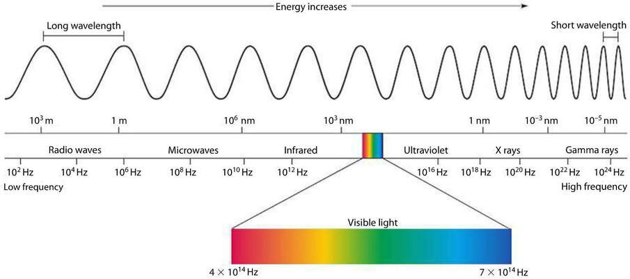
The parts on the left have the longest wavelength and lowest frequency (radio waves), and the parts on the right have the shortest wavelength and highest frequency (gamma rays). As we go from left to right, the wavelengths get shorter and the frequencies increase.
The effects that the different parts have on the body depend on how much energy the waves are transferring. The dangers associated with electromagnetic waves increases as the wavelengths get shorter and the frequencies increase.
Low frequency waves don’t transfer much energy and tend to pass through soft tissue without being absorbed; radio waves have no known damaging effects on the body.
High frequency waves cause more damage as they transfer more energy. Ultraviolet waves, X-rays and gamma rays are all types of ionising radiation. Ionising means that these waves can add or remove electrons from molecules, resulting in the molecules becoming electrically charged (ions). This can result in cells mutating, which can lead to cancer. Here are the effects of ultraviolet waves, X-rays and gamma rays:
- Ultraviolet waves can cause skin cells to become damaged (sun burn), skin cancer, skin to age and can damage your eyes (potentially even blindness).
- X-rays and gamma rays can cause genes to mutate, which can potentially lead to cancer. X-rays and gamma rays can also cause cells to die.
Radiation Dose
Radiation dose measures the risk of harm when the body is exposed to different types of ionising radiation. Radiation dose is measured in Sieverts (Sv). As radiation dose is generally very small, we tend to measure radiation dose in millisieverts (mSv) where 1 Sv is 1,000 mSv.
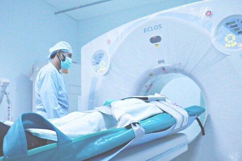
Before we use ultraviolet waves, X-rays and gamma rays for medical procedures, we need to weigh up the benefits and costs of their use. For example, let’s suppose that an individual has been in a car crash and has potentially broken his arm. The doctors treating this individual will take an X-ray of the arm to see whether the arm is or is not broken. The benefits of finding any broken bones/ other issues from the X-ray massively outweighs the risk of exposing the injured individual to a small amount of radiation during the X-ray. We can also reduce the risk associated with the X-ray by only exposing parts of the body that need to be exposed to X-rays. For our individual in the car crash, his arm was potentially broken, so we just expose his arm to the X-rays. Also, the individual having the X-ray may be given a metal plate to cover parts of the body where gametes (sex cells) are stored (the testes in males and the ovaries in females). Furthermore, the radiographer in charge of the X-ray machine will either stand behind a screen or leave the room when the X-ray is taking place.
We would also need to compare the costs and benefits of using medical tracers that produce gamma rays to find the location of an internal bleed inside an individual. The costs of using medical tracers that give off gamma rays is that the radiation dose that the individual is exposed to is quite high, which may lead to cells mutating and the individual developing cancer. However, the medical tracers will give doctors a good level of detail about the location and size of the potential internal bleed. This allows doctors to make a very informed decision about the best treatment to solve the internal bleed, thus meaning that the treatment may be more effective. If the medical tracers weren’t used to find the internal bleed, there would be an increased chance of the individual dying or having a reduced quality of life. Therefore, for this case, the benefits of using the medical tracers will outweigh the costs/ risks of using the tracers. However, we would need to consider the benefits and costs of other methods that we could use to diagnose the internal bleed.
Radiation dose measures the risk of harm when the body is exposed to different types of ionising radiation. Radiation dose is measured in Sieverts (Sv). As radiation dose is generally very small, we tend to measure radiation dose in millisieverts (mSv) where 1 Sv is 1,000 mSv.
Before we use ultraviolet waves, X-rays and gamma rays for medical procedures, we need to weigh up the benefits and costs of their use. For example, let’s suppose that an individual has been in a car crash and has potentially broken his arm. The doctors treating this individual will take an X-ray of the arm to see whether the arm is or is not broken. The benefits of finding any broken bones/ other issues from the X-ray massively outweighs the risk of exposing the injured individual to a small amount of radiation during the X-ray. We can also reduce the risk associated with the X-ray by only exposing parts of the body that need to be exposed to X-rays. For our individual in the car crash, his arm was potentially broken, so we just expose his arm to the X-rays. Also, the individual having the X-ray may be given a metal plate to cover parts of the body where gametes (sex cells) are stored (the testes in males and the ovaries in females). Furthermore, the radiographer in charge of the X-ray machine will either stand behind a screen or leave the room when the X-ray is taking place.
We would also need to compare the costs and benefits of using medical tracers that produce gamma rays to find the location of an internal bleed inside an individual. The costs of using medical tracers that give off gamma rays is that the radiation dose that the individual is exposed to is quite high, which may lead to cells mutating and the individual developing cancer. However, the medical tracers will give doctors a good level of detail about the location and size of the potential internal bleed. This allows doctors to make a very informed decision about the best treatment to solve the internal bleed, thus meaning that the treatment may be more effective. If the medical tracers weren’t used to find the internal bleed, there would be an increased chance of the individual dying or having a reduced quality of life. Therefore, for this case, the benefits of using the medical tracers will outweigh the costs/ risks of using the tracers. However, we would need to consider the benefits and costs of other methods that we could use to diagnose the internal bleed.
|
|
Comparing Risk
You may be asked in the exam to compare the radiation doses for different medical procedures.
Question
The radiation dose for a CT scan of the brain is 1.6 mSv and the radiation dose for a PET scan is 22.7 mSv (PET is a medical tracer technique). How many times greater is the radiation dose from a PET scan compared to the radiation dose from a CT scan of the brain; give your answer to the nearest integer.
We can find this out by dividing the radiation dose from the PET scan (22.7 mSv) by the radiation dose from the CT scan of the brain (1.6 mSv).

This tells us that a PET scan gives a radiation dose that is 14 times greater than the dose from a CT scan of the brain.